Start with a disc. Or a folder.
Open DICOMDIR media or scan a folder and its subfolders. MedView groups available images, series and DICOM Structured Reports for browsing.
Background scanning · Multi-patient navigation

THE MEDVIEW DESKTOP WORKSPACE
Open the study. Compare the series. Explore it in 3D. Your images, measurements and reports, together in one DICOM viewer.
Windows x64 · 15-day trial for new installations
A CLOSER LOOK
Make space for comparison, reconstruction or the report. Explore these real MedView screens.
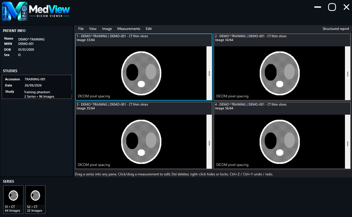
Drag a series into a pane. Choose 1, 2, 4 or 8 views, then navigate independently or link image and frame scrolling.
FROM IMPORT TO INSIGHT
Open DICOMDIR media or scan a folder and its subfolders. MedView groups available images, series and DICOM Structured Reports for browsing.
Background scanning · Multi-patient navigationUse up to eight panes, drag different series into each view, and keep window settings independent. Scroll by mouse, keyboard or the side scrollbar.
1 / 2 / 4 / 8 panes · Optional linked scrollingAdjust window and level, choose presets, fit or view at 1:1. Zoom, pan, invert, flip and rotate without changing the source DICOM files.
Window/level presets · Multiframe navigationSwitch tools without losing your work. Select drawings on the image, move them, resize their handles, reposition labels, or hide and lock individual annotations.
Delete · Undo/redo · Copy/paste · Local savingHover to preview a DICOM Structured Report, click to pin it, then resize its panel. The image workspace adjusts to the space available.
Study-linked reports · Multiple SR documentsSave 3D workspaces and view bookmarks. Export PNG captures, derived DICOM Secondary Captures, or a 360° rotation as an AVI video.
Workspace files · View bookmarks · Image & video exportA DIFFERENT PERSPECTIVE
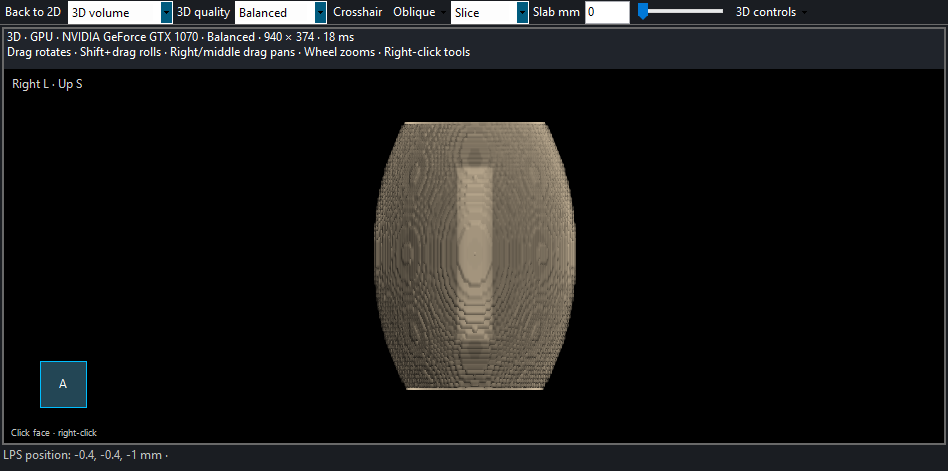
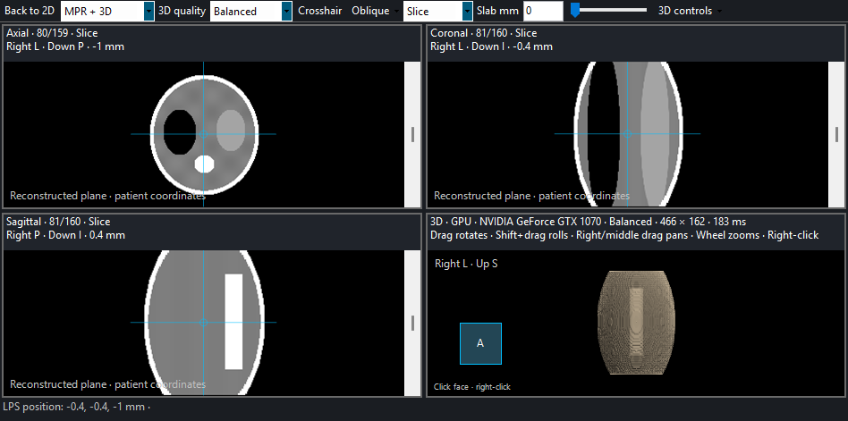
Reconstruct compatible CT and MR series. Move between linked planes and an interactive volume without leaving the workspace.
Axial, coronal and sagittal reconstruction with shared crosshairs. Rotate oblique planes, set slab thickness and choose MIP, MinIP or Average.
Fast, Balanced and High quality modes. Responsive previews refine after interaction, with CPU fallback and memory-aware loading for larger volumes.
Drag crop handles, add clipping planes, or keep and remove drawn regions. Restore cuts and undo edits while retaining the source data.
APPEARANCE
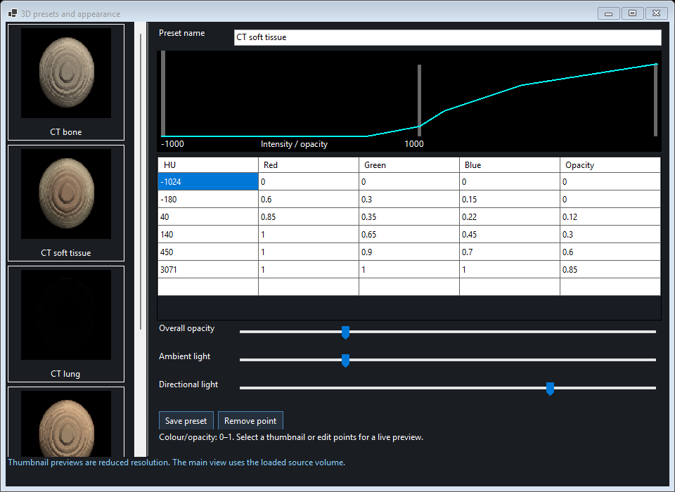
Thumbnail presets for CT bone, soft tissue, lung, contrast vessels and abdomen, plus MR intensity presets. Adjust colour, opacity and lighting, then save your own.
TISSUE TOOLS
Threshold and connected-region selection, named colour layers, MPR paint/erase brushes and volume estimates. Assisted bone and seeded object removal keep correction in your hands.
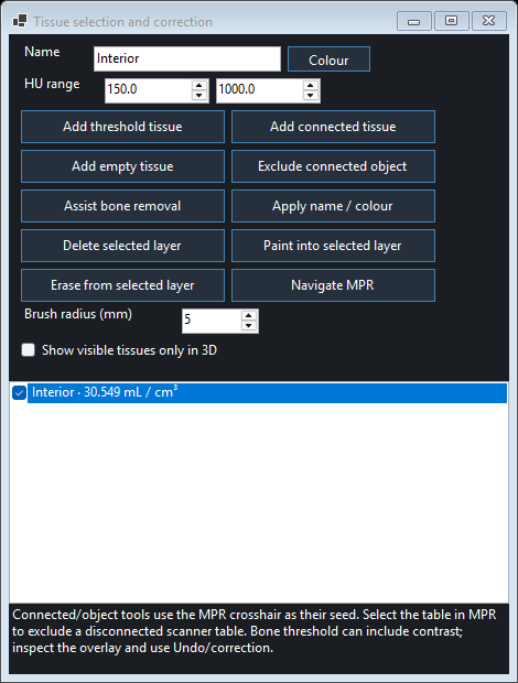
3D requires a compatible monochrome CT/MR series with reliable spatial metadata. Tissue tools provide assisted selection and voxel-based volume estimates; they do not automatically identify organs.
MEASURE, THEN REFINE
Create a measurement once. Keep adjusting it as you review.
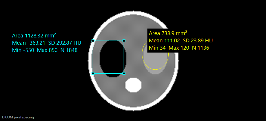
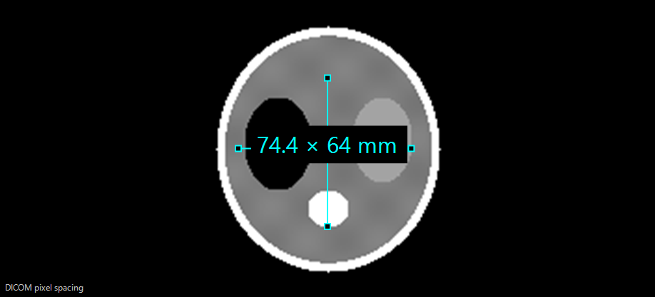
Physical measurements use available DICOM pixel spacing. Choose mm, cm or automatic units, and calibrate a known length when needed. Missing calibration is clearly shown in pixels.
Distance, polyline, angle, Cobb angle, bidirectional/perpendicular, deviation and length ratio.
Rectangle, ellipse and polygon ROI, plus a pixel probe. Statistics include mean, minimum, maximum and standard deviation.
Arrow/text, pencil, movable labels, hide/lock, bulk clear, copy/paste and undo/redo.
A CONNECTED WORKFLOW
Choose a DICOMDIR or scan a folder and its subfolders.
Drop series into panes and choose independent or linked scrolling.
Measure, read the report, and reconstruct compatible CT/MR data.
Revisit saved annotations and workspaces, or export a view.
CHOOSE YOUR WORKFLOW
MedView supports both bundled viewing and a separately licensed desktop application. Contact BOSS-InfoTech for the edition that fits your deployment.
BUNDLED EDITION
For supported media created through the BOSS-InfoTech disc publishing workflow.
Capabilities depend on the bundled build. The screenshots and advanced features on this page describe the current desktop application.
Discuss disc deploymentDESKTOP EDITION
15-day trialFor broader DICOMDIR and folder workflows on your Windows workstation.
License details and renewal checks are available inside File → License.
YOUR NEXT VIEW STARTS HERE
Download the Windows application and explore the 15-day trial. For activation or renewal, contact BOSS-InfoTech with the product key and machine ID shown in the app.
New installations register the trial silently when the licensing service is available.
GOOD TO KNOW
Questions about licensing or your imaging workflow?
Talk to BOSS-InfoTechNo. The desktop app can scan a selected folder and all subfolders, including extensionless DICOM files. Available images and DICOM Structured Reports are grouped into studies and series. Keep the source drive connected while viewing.
Yes. Select a compatible series, open View → 3D volume, then choose 3D controls → Presets and appearance. Click a thumbnail to apply it. CT presets include bone, soft tissue, lung, contrast vessels and abdomen; MR has separate intensity presets.
Compatible monochrome CT/MR series with consistent spatial metadata, including supported enhanced multiframe data. Missing geometry, mixed slice positions or large unexplained gaps may prevent reconstruction. A single projection image does not form a 3D volume.
No. Completed drawings remain and can be selected, moved, resized, hidden, locked or deleted. Annotations save locally and are associated with the source image/frame or exact MPR plane. They are not automatically embedded in DICOM files or transferred with a study.
The current decoder covers common uncompressed formats, RLE, JPEG, JPEG-LS, JPEG 2000 and HT-JPEG 2000 variants. Coverage is not universal: DICOM video, some retired/private encodings and unsupported object types are outside the current implementation.
The report panel displays DICOM Structured Reports associated with the study. Standalone or encapsulated PDF reports are not displayed as SR. The 3D workspace exports PNG, rendered RGB DICOM Secondary Capture and rotation AVI, and saves .mv3d workspaces. Exported RGB captures are display images, not quantitative CT/MR source voxels.
PACS transfer is not available in the current build. You can review supported exported DICOM studies locally; network query/retrieve and study transmission are not advertised as current features.
New installations receive a 15-day trial. Open File → License for the product key, machine ID, license type and expiry. Provide those identifiers to BOSS-InfoTech for activation or renewal, then use Check license. An unexpired cached license can support offline startup, subject to its validation status.